Lattice Degeneration is a thinning of the Retina that happens over time

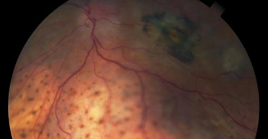
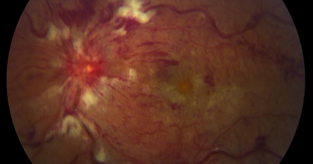
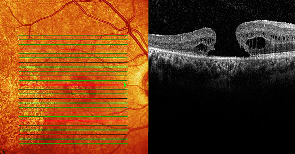
Lattice Degeneration is a thinning of the Retina that happens over time. Although 10% of the population has Lattice Degeneration, most will not have any symptoms or loss of vision as a result. Rarely, Lattice Degeneration may lead to Retinal Detachment. While doctors aren’t certain about what causes Lattice Degeneration, it’s most common in people with myopia, or nearsightedness, and in people with diseases like Stickler or Marfan syndrome. Although it is not genetically passed down from parents, those with a family history are more likely to also contract it. Lattice Degeneration doesn’t typically have symptoms, but because the Retina is thinner it is more subject to holes, tears, and breaking, which can in turn lead to Retinal Detachment, which can cause blindness if not treated immediately. Learn more about Lattice Degeneration at https://bit.ly/3gE9BM6

Retinoblastoma is a rare type of eye cancer that usually develops in early childhood

Retinoblastoma is a rare type of eye cancer that usually develops in early childhood, typically before age 5. This form of cancer develops in the Retina, and typically affects only one eye, although one out of three children with Retinoblastoma eventually develop the cancer in both eyes. The most common first sign of Retinoblastoma is a visible white spot in the pupil, also called a “cat’s eye reflex,” that is noticeable in dim light or in photographs taken with a flash. Other symptoms of Retinoblastoma include crossed eyes, or eyes that don’t point in the same direction. Retinoblastoma is often curable when diagnosed early, but if not treated promptly it can spread beyond the eye to other parts of the body. Learn how one mother’s intuition saved her son’s eye, and his life at http://bit.ly/39V3Wun.

Those who experience chronic red, itchy eyes may be suffering from Pinguecula, or Pterygium

Those who experience chronic red, itchy eyes may be suffering from Pinguecula, or Pterygium. A Pinguecula is a raised, yellowing deposit of protein and fat that develops in the thin membrane that covers the white part of the eye. A Pterygium is a growth of fleshy tissue containing blood vessels that often starts as a Pinguecula, but can grow and affect vision. Both Pinguecula and Pterygium are thought to be caused by excessive sun and wind exposure. Pterygium is also known as “Surfer’s Eye” or “Farmer’s Eye”. While these growths are non-cancerous, they can become irritated and inflamed, and can be accompanied by burning, itching, and tearing. To prevent Pinguecula and Pterygium, avoid excessive sun exposure and wear sunglasses that block UV rays. While these conditions tend not to go away on their own, they may not require removal unless they impact vision. Learn more about “Surfer’s Eye” at http://bit.ly/3skeVXv, and be sure to contact a Retinologist at High Country Macula to discuss any eye irritation that affects vision.

A breakthrough advancement in the development of a cell therapy to treat blindness

Scientists have made a breakthrough advancement in the development of a cell therapy to treat blindness. A recent study showed that Retinal cells from a human cadaver eye can survive when transplanted into a primate model. The Retinal Pigment Epithelium, or RPE, is a layer of pigmented cells in the Retina which functions as a barrier and regulator to maintain vision. RPE dysfunction can lead to a multitude of eye disorders, including Age-related Macular Degeneration. The study found that RPE patches from donated adult cadaver eyes could partially replace the function of the RPE in the Macula of a non-human primate. Although the study suggests that donor RPE is safe to transplant, further studies are necessary to determine whether RPE stem cells from donated eyes could restore vision in human patients. Learn more about this fascinating study at http://bit.ly/3aeDYnl


Retinitis Pigmentosa is a group of rare genetic disorders that involve a breakdown and loss of cells in the Retina

Retinitis Pigmentosa (RP) is a group of rare genetic disorders that involve a breakdown and loss of cells in the Retina. RP is an inherited disorder that results from gene mutations that damage photoreceptors in the Retina. There are two types of photoreceptors – Rods, which allow us to see in dim and dark light, and Cones, which allow us to perceive fine visual detail and color. Symptoms of RP typically appear in childhood. Children may have difficulty getting around in the dark, and as their visual field becomes restricted, they often trip over things or appear clumsy. RP progression varies greatly from person to person. Some people experience a restricted visual field into their 50s, while others experience significant vision loss in early adulthood. Eventually, individuals with RP will lose most of their sight.
Share this with others to help to preserve vision and visit both http://bit.ly/2CXyGfF and highcountrymacula.com to learn more.

What is Vitreomacular Traction?

What is Vitreomacular Traction (VMT)? The middle of the eye is filled with a substance called Vitreous. In a young, healthy eye, this gel-like substance is firmly attached to the Retina and Macula by millions of microscopic fibers. As we age, or as a result of eye disease, the Vitreous shrinks and pulls away from the Retina, and over time completely separates, which is normal and happens to most people by age 70. But in some people, the Vitreous doesn’t detach completely, and remains partly stuck to the Macula at the center of the Retina. The Vitreous then pulls and tugs on the Retina, causing VMT, which can create vision loss if not treated. If VMT is mild and doesn’t impact vision, treatment may not be needed, and may only require home monitoring with an Amsler Grid. Severe cases can lead to vision threatening conditions such as Macular hole, when tugging of the vitreous creates a hole in the Macula, or Macular pucker, when macular scar tissue builds up and distorts vision. If ones vision is impaired, it is urgent to contact a Retinologist at High Country Macula to preserve vision. Learn more at highcountrymacula.com and at https://bit.ly/36icbSa

Although eye cancer is rare, the most common form is Choroidal Melanoma

Although eye cancer is rare, the most common form is Choroidal Melanoma, which originates in the Choroid, a dense layer of vessels that supplies blood to the Retina. In its early stages the cancer doesn’t interfere with vision, but later it may cause blurred vision or Retinal Detachment, with symptoms such as flashes of light or the illusion of a veil across the visual field. Regular eye exams are important, as early diagnosis of Choroidal Melanoma can be treated with laser or radiation treatments which may preserve vision and save the eye. Contact your Retinologist at High Country Macula to learn more.

Optic Neuritis occurs when the optic nerve that transmits visual information from the Retina to the brain becomes inflamed

Optic Neuritis occurs when the optic nerve that transmits visual information from the Retina to the brain becomes inflamed. When inflammation causes damage to the myelin, or protective sheath surrounding the nerve, the resulting neuritis can lead to blurring, blind spots, or complete loss of vision. It may also lead to loss of color vision, or pain when you move one or both eyes. Optic Neuritis can occur in anyone, but it usually occurs in adults younger than 45, and effects more women than men.
During an eye exam, your Ophthalmologist will check for signs of Optic Neuritis by conducting tests to evaluate whether you have reduced vision, a visual field test to determine if you have an enlarged blind spot, and Optical Coherence Thermography (OCT) to determine if there has been damage to the optic nerve fibers. Regular eye exams by your Retinologist at High Macula are imperative. If identified and treated early, about 95% of people Optic Neuritis will recover much of their vision within 6 months of onset. Learn more about Optic Neuritis at https://bit.ly/2YDvH81 .

Endophthalmitis is an infection of the tissue or fluids inside the eyeball

Endophthalmitis is an infection of the tissue or fluids inside the eyeball. The most common type of Endophthalmitis is Exogenous Endophthalmitis, when the source of the infection comes from outside the body. Bacteria may get inside the eye during surgery, an injection to the eyeball, or an eye injury. Puncture wounds to the eye are the most likely cause of Endophthalmitis. The other type of Endophthalmitis is Endogenous Endophthalmitis, which starts as an infection in another part of the body, for example a urinary tract or blood infection, which then spreads to the eye. If you or a loved one suffers eye pain that gets worse after surgery, an injection or injury, or if you experience discharge from your eye, be sure to contact your Retinologist at High Country Macula, immediately. Endophthalmitis is an urgent medical emergency, and can cause blindness if not treated quickly. Learn more at http://bit.ly/37vn8xO

If you suffer from Atherosclerosis, you may also be at risk for Retinal Vascular Occlusion

If you or a family member suffers from Atherosclerosis, you may also be at risk for Retinal Vascular Occlusion. Your Retina requires a constant supply of blood to make sure your cells receive enough nutrients and oxygen. If a blood vessel carrying blood to or from the Retina becomes blocked, or a clot occurs, you may experience occlusion. The primary symptom of Retinal Vascular Occlusion is a sudden change in vision, either sudden blurriness or partial or complete loss of vision, typically in just one eye. Be sure to immediately contact your Retinologist at High Country Macula should you unexpectedly experience blurriness or a loss of vision.